
Education & Resources
Renal Physicians
about | home
| |
|
|
Education & Resources Renal Physicians |
communication | vision about | home |
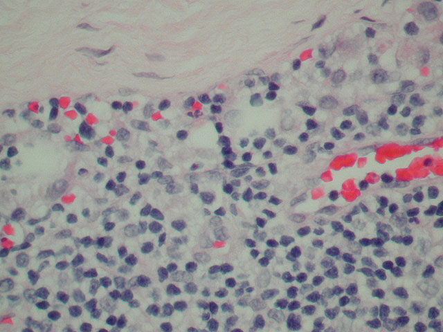
This is an example of keratin positive cells in a lymph node from a patient with breast cancer; no diagnostic metastatic carcinoma was seen with H&E stains of this sentinel lymph node. The first picture on the left shows the subcapsular sinus histiocytes in an H&E stained level. The middle picture is a scanning view of the keratin stain with single, positive staining cells in the subcapsular sinus. The picture on the right is a high power, showing keratin staining of cytoplasm and staining of the nucleus with a counterstain indicating it is a cell and not artifact.
To View Original Image Click on Thumbnail
 |
![Single Keratin Pos cells[1].jpg (100416 bytes)](http://www.cybernephrology.org/education/Koelliker/Images/Single_Keratin_Pos_cells1.jpg) |
![Single Keratin Pos[1].jpg (116216 bytes)](http://www.cybernephrology.org/education/Koelliker/Images/Single_Keratin_Pos1.jpg) |
| Copyright © 2000-2007 cyberNephrologyTM
All rights reserved. Last Modified: Thursday March 08, 2007 05:21:14 PM |
info@cybernephrology.org |