
Education & Resources
Renal Physicians
about | home
| |
|
|
Education & Resources Renal Physicians |
communication | vision about | home |
Dear Nephrolers':
I now have the capability of transferring images directly from my fluoroscopy unit to
the computer. I thought I would post an interesting case I just had. If people
like this, I can post other cases and if anyone has a particular interest I can post an
example when I get one (I cannot transfer my old film library as they are kept only on cut
film). Just a warning, these images take a large amount of space if you elect to download
them.
This is a four year old radial artery fistula which presented to our LifeLine Vascular Lab
clotted for less than 48 hrs.
In order to access the anastomotic area of the fistula I had to go through the brachial
artery.
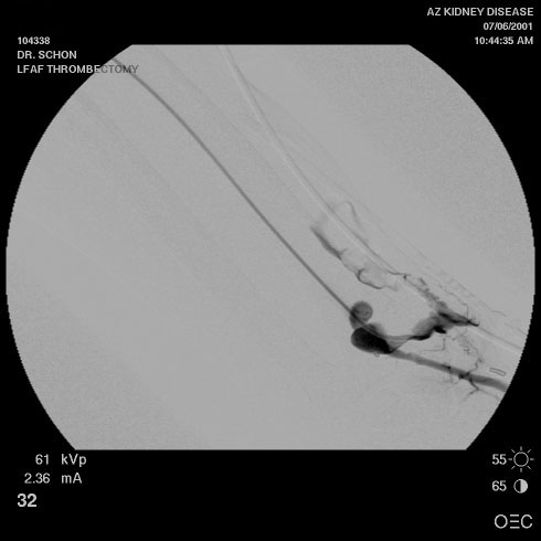
The first image is of the anastomosis from the arterial side. To the left you see the artery. The beaded portion near the wrist (the clips mark the anastomosis) is the stenosis and it is in the radial artery itself extending into the anastomosis. What the film demonstrates is the clot which extended from the anastomosis to the elbow.
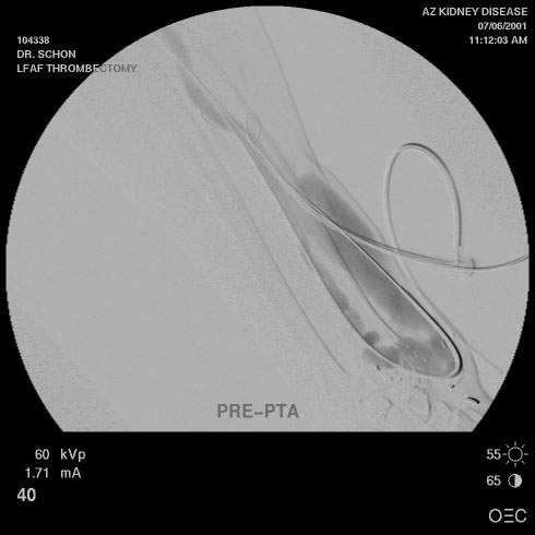
The second image demonstrates the fistula after thrombolysis with a total dose of 1.25 mg of tPA. The clot is gone and the anatomy of the forearm portion of the fistula is clear.
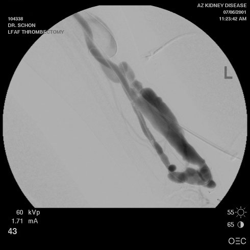
The third image is of the fistula after PTA of the radial arterial stenosis to 6 mm
(the normal portion of radial artery measured 5mm prior to PTA). The post arterial
anastomotic section of the fistula (PAAS) distal to the anastomosis is about 6 mm and the
narrowest area of the fistula itself distal to this is 8mm so it required no further
dilatation.
Please give me feed back if you find this valuable.
Thanks
Donald Schon, MD, FACP
LifeLine Vascular Lab
Phoenix, AZ
To View Original Image Click on Thumbnail
 |
 |
 |
| Copyright © 2000-2007 cyberNephrologyTM
All rights reserved. Last Modified: Thursday March 08, 2007 05:21:13 PM |
info@cybernephrology.org |