
Education & Resources
Renal Physicians
about | home
| |
|
|
Education & Resources Renal Physicians |
communication | vision about | home |
Dear Nephrolers':
There has been a lot of recent discussion of fibrin sheaths and SVC clot.
Therefore I thought that people might like to see some examples:
Image 1 "svc Clot" is a clot at the end of a fibrin sheath. The catheter
has been removed already.
Image 2 "post pta" is after I have mascerated the clot by angioplasty
with a 14 mm balloon. Because of the small residual clot I have recommended that the
patient be anticoagulated for a couple of months.
Image 3 is of a double fibrin sheath from a twin cath system. Please note
that the catheter has been withdrawn to the top of the clavicle and what looks like two
catheter lumens are just the sheaths.
Image 4 "retrograde filing" is fascinating. The IJ has been separately
punctured low in the neck (see the micropuncture dilator to the left of the sheath) and
the injection of contrast is through this. A Tessio lumen has been withdrawn to the
level of the upper neck (the other lumen fell out which is why the referral and I replaced
them both with a
SchonCath placed lower in the IJ). The sheath between the Innominate and the
catheter lumen is filling retrograde from the innominate vein. Thus you clearly see the
junction of the sheath with the innominate, and the sheath with the partially withdrawn
Tessio lumen.
I hope that these are interesting and helpful.
Thanks
Donald Schon, MD, FACP
LifeLine Vascular Lab
Phoenix, AZ
(To View Original Image Click on Thumbnail)
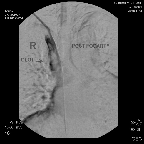 Image 1 |
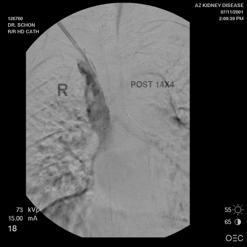 Image 2 |
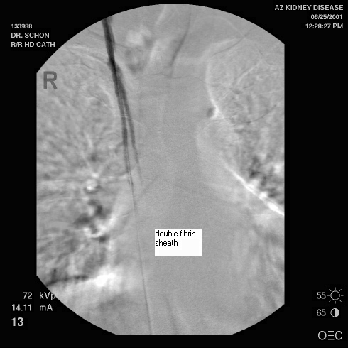 Image 3 |
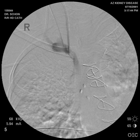 Image 4 |
| Copyright © 2000-2007 cyberNephrologyTM
All rights reserved. Last Modified: Thursday March 08, 2007 05:21:10 PM |
info@cybernephrology.org |